

Oesophagus Practical Case.
A 44-year-old man that suffered from an oesophageal ulcer two years ago as the result of vomiting. His doctor diagnosed him with Boerhaave Syndrome.
In the photograph of his left iris, we can see how in the Topographic Area (TA) of the oesophagus we find a “Lacuna” sign with a very defined relief, where we can observe depth. This indicates a chronic weakness where, in a specific moment, vomiting had led to a spontaneous rupture of the oesophagus that came close to killing the man.
|
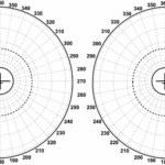
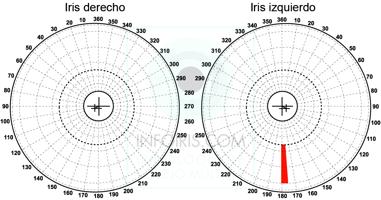
The Topographic Area (TA) that conveys changes to the oesophagus is found in the left iris between 176º and 181º in crowns 4, 5 and 6. It corresponds to the area coloured in red. |
 |

Left iris. 44-year-old man.
When we find the “Lacuna” sign with relief where we can see the bottom, it is a sign of a chronic weakness and greater degenerative risk. In this case, the oesophagus has suffered from the aggressive action of gastric juices over a long period of time. Normally, we often find this type of sign, or very similar signs, with Barrett’s Oesophagus, as we will see in some cases.
Here, several factors have all played a role: a very weak oesophagus as the result of aggression to tissues that were in themselves weak, whereby an episode of vomiting attacked that area, causing a rupture of the oesophagus. The erosion of the gastric juices and the stress produced, was what caused the rupture.
We have photographs of before and after the incident. Strangely, this person was not aware of having suffered from acidity. However, the “Lacuna” sign already indicated a very marked weakness to those tissues, that over the course of time, ended up giving way to the strain that was greater than their ability to resist.
(The rest of the image of the iris and the interpretation of it is reserved for courses and study materials that we will gradually publish.)
Importance of the observation technique.

In this instance I wanted to make the most of the same iris and show photographs of it at the same time using the same equipment, but with a different angle of light so that we can appreciate how different details stand out.
| More information about oesophageal disorder on pages 303 to 306 of the book “Principios y fundamentos de la Iridología de Reflejo Múltiple”. Editorial infoiris |
© Multiple Reflex Iridology. All rights reserved. © Javier Echavarren Otín. 2017


