

The iris changes over the years. This is a reality that despite the existing evidence, some dare to deny. My biggest surprise is when I find iridologists who support this denial. Surely they do not have a large database that collects photographs of irises over the years, because that is the only way to check if it occurs or not.
In my case, I’ve been making and archiving iris photographs of thousands of people for 15 years that allow me to document this fact. In some photographs you can see obvious changes in an indisputable and unquestionable way.
Another reason to make this article is to expose and reflect the complexity of some of the changes that occur in the iris. For this reason, I have chosen as an example for this article a part of an iris in which different changes occur. Some more obvious and others more subtle, but no less important in the interpretation of the information it gives us to Iridology.
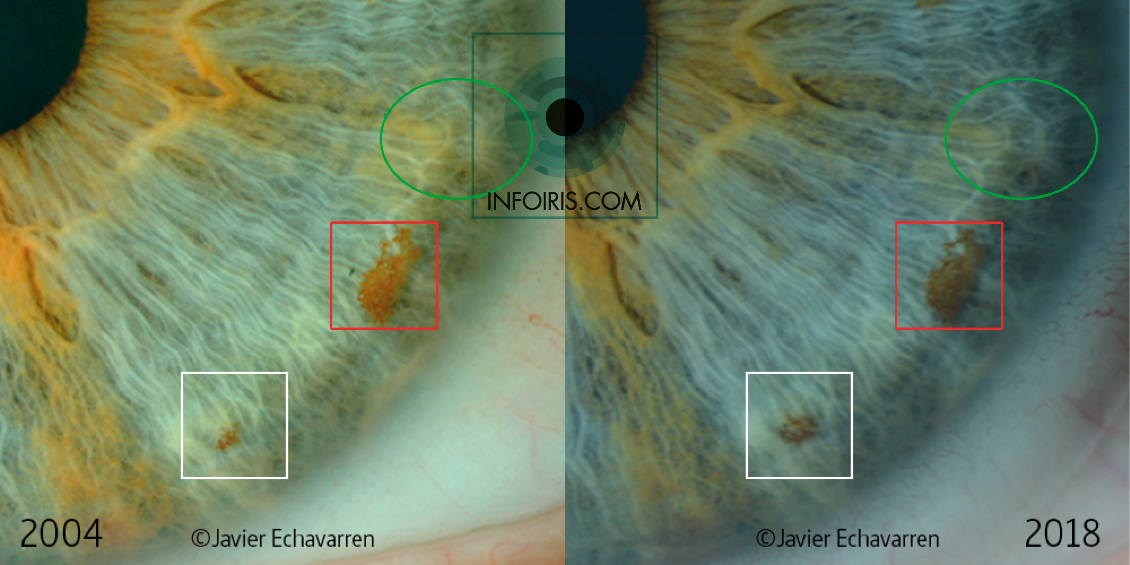
We are going to observe a part of Sector 2 (S2) of a left iris of a 48-year-old woman (2018). We will observe photographs taken in 2004 and 2018, so we will have a difference of 14 years.
The iridian signs may undergo changes over the years for different reasons. It depends on the characteristics of the sign itself, the factors or causes that have acted in its genesis and development, and finally the incidence of aging or deterioration of the tissues or structures of the eye itself. This last factor is not relevant in this particular case. Consequently, we must take into account the different factors that are influencing the characteristics of the iris for a more accurate assessment and analysis.
I wanted to highlight several areas where we can see changes in the iridian signs manifested in the iris tissue.
I am used to finding comments of all kinds. Some will tell me that there are more changes in the iris tissue, because it really is. And instead, I will also find comments that will only admit the changes collected in the blank boxed area due to its overwhelming evidence. All this will depend on the degree of knowledge, experience, skill and attitude that the person making the contribution has.
For that reason I will start in the highlighted areas. I have included several forms of presentation of the photographs of 2004 and 2018. In two cases they can be observed superimposed with the application of the Parallax effect so that anyone can move the timeline that separates the two photographs to their liking and observe the changes produced in Any part of the picture. I found one of the best tools to compare the photographs.
Also included is a video that shows an example of the use of the application of the Parallax effect.
I have to mention something obvious about the characteristics of the photographs. Despite using the same Slit Lamp (Topcon SL8Z), over the years I have changed camera so the light conditions setting is not the same. Consequently, a change in the general color of the iris color that does not correspond to any change in the surface of the iris is evident. This detail is exposed and taken into account with respect to the perception of the rest of the changes in the iris. This is one of the reasons why I often rule out certain examples of changes that may be doubtful because I have not achieved the same light conditions. I mention it for two reasons. First, because it is necessary to declare this factor, and second, because it is a very frequent fact and very difficult to avoid.


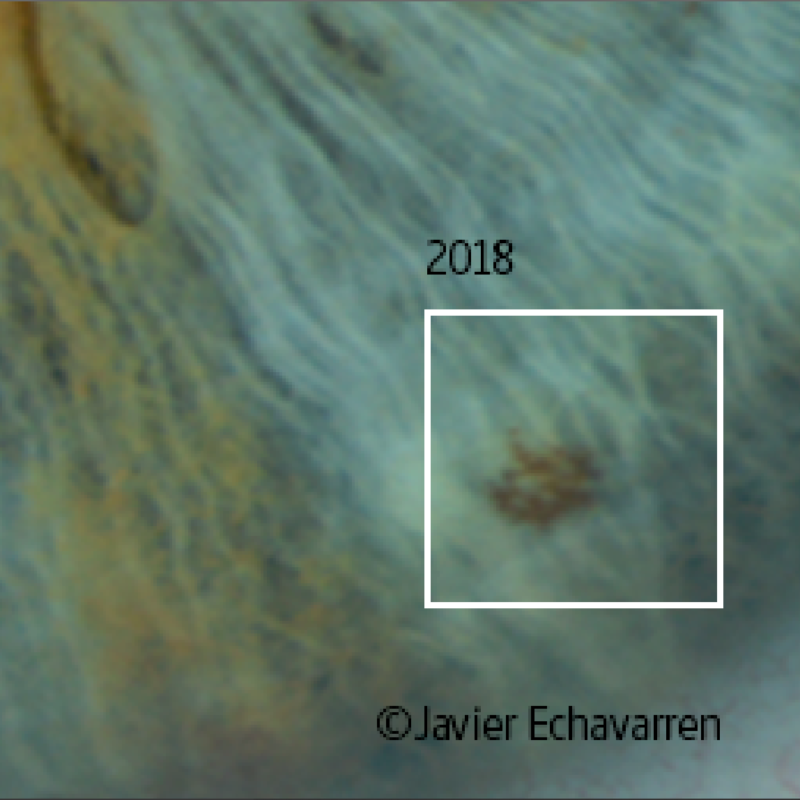
The increase in size of a Pigmentation located inside a Lacuna is clearly visible in the blank boxed area.
You can find this extended area with the application of the Parallax effect. By manually moving the timeline it can be seen that not only does the Pigmentation change in size, but on the left side of the white square where the iridian stroma has another color (orange and brown) in 2018 there is a lower density of the Orange Pigmentation.
In this article I do not enter into the assessment and interpretation of the iridian signs, that information is reserved for specific courses and materials.
In the red boxed area we observe a Stain of an important size on a Transversal sign. This Stain is where subtle changes occur. Some of them could be said without being very wrong that there are almost no changes and that the appreciation of that change is due to the different light conditions.
Although, I have already mentioned the difficulty of always achieving the same light conditions, other factors must be taken into account. It is important to make annotations at the time of taking the picture. Record if there has been any change or if the actual color tones do not match those reflected in the photograph. A fact that I take into account when starting the first photographs of each session, contrasting the colors of the iris with the photographs on the screen to adjust the settings of the following photographs.
For this reason and because after the photographs I start comparing with previous photos, I record in my patient notes if the color tones of the iris collected in the photographs fit the real ones or not.
In this case my records pick up that change in the general color of the iris, but also confirm the slight darkening of the Stain. In some areas there is an increase in color density. There are also subtle changes in the upper part where a hole appears through which the Transversal is seen that crosses through its lower face.
In the area indicated within the green circumference we find several Transversals. One that starts from the Topographic Area (TA) of the spleen and ascends to the TA of the lower lobe of the left lung. This first branches into two branches within the green circumference. The second Transversal is vascularized.
There is a white stroma that stands across the center of the circle horizontally. On this white stroma in the part that corresponds to the left side of the circle, it can be seen in a blur like an orange Pigmentation cloud that underlines the upper part of the mentioned stroma. This orange coloration that especially stands out in this elongated shape, over the years it has blurred, reducing its density.

All these changes mentioned are another example that demonstrates how the appearance of the iris undergoes changes over time. Then I leave links to other examples that confirm this fact, but as it is a topic that can be talked about a lot, I will continue making contributions and enriching with more graphic documents.
On the web you can find other articles related to changes in the iris and evolution of the iridian signs by selecting the link of the tags: “Changes in the irises”, “New iridian sign”.
Some of the featured articles are as follows:
– New Stain in the Topographic Area of the spleen.
– New pigmentation in the Topographic Area of the cardia.
– Video of the evolution of an iris.
– Video of the evolution of a Stain in the TA of the left lung.
– Increase in a Pigmentation on the iris (TA of the liver).
– Radical change of iris color.
Some of the videos that show the changes that can be seen on the surface of the iris can be found on Facebook, Instagram or on my YouTube channel.
© Multiple Reflex Iridology. All rights reserved. © Javier Echavarren Otín. 2019
More from my site
 Pigmentation increased in 11 years
 Case 1. Increased “Stain” in the Topographic Area (TA) of the left lung.
 New micropigmentations in the Topographic Area of the left lung
 New Stain in the Topographic Area of the spleen.
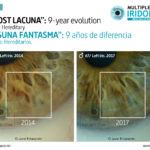 Case 18. Ghost Lacuna
 Video of the evolution of a Stain Sign in the Topographic Area of the liver